Established in September 2017 and headquartered in Northern Holland, by setting the extension researcher plan together with Dutch universities, HyperLab has established itself as a trusted provider of robust, end-to-end solutions for the spatial biology field, supporting both theoretical exploitations and clinical professionals in major direction. Our core mission is to engineer advanced hardware and integrated systems that serve as the foundational backbone for scientific discovery, enabling researchers to uncover deeper biological insights with precision and reliability.
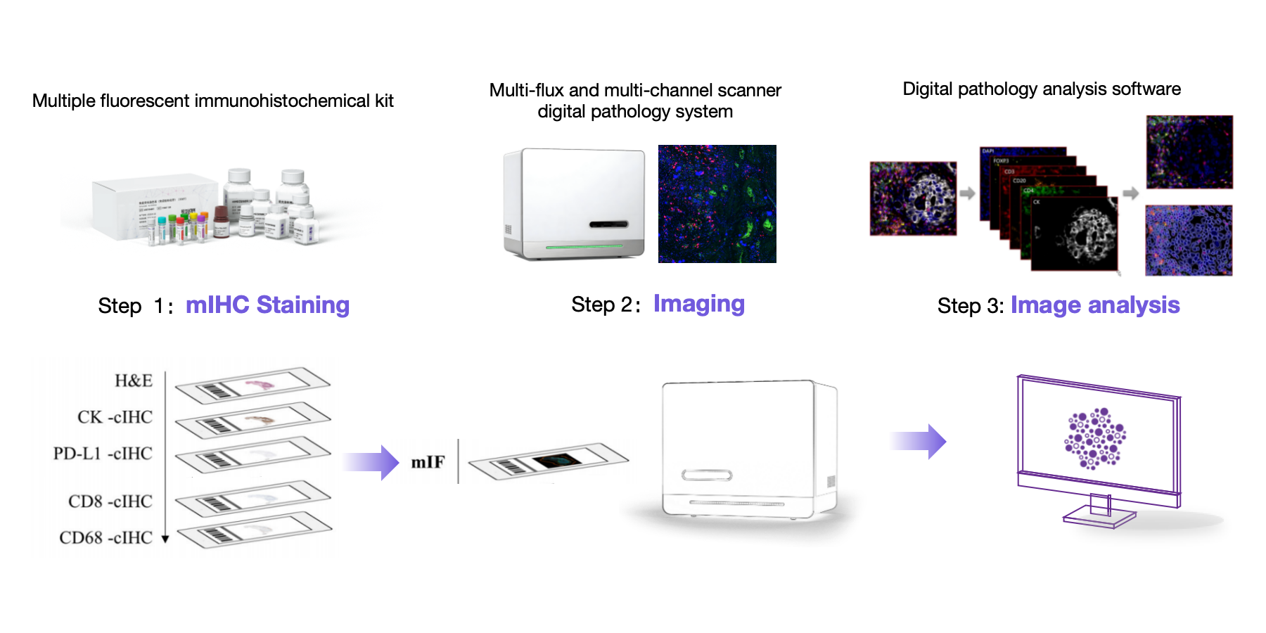
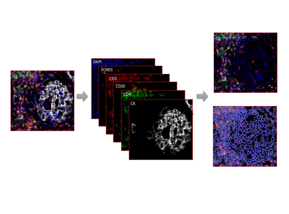
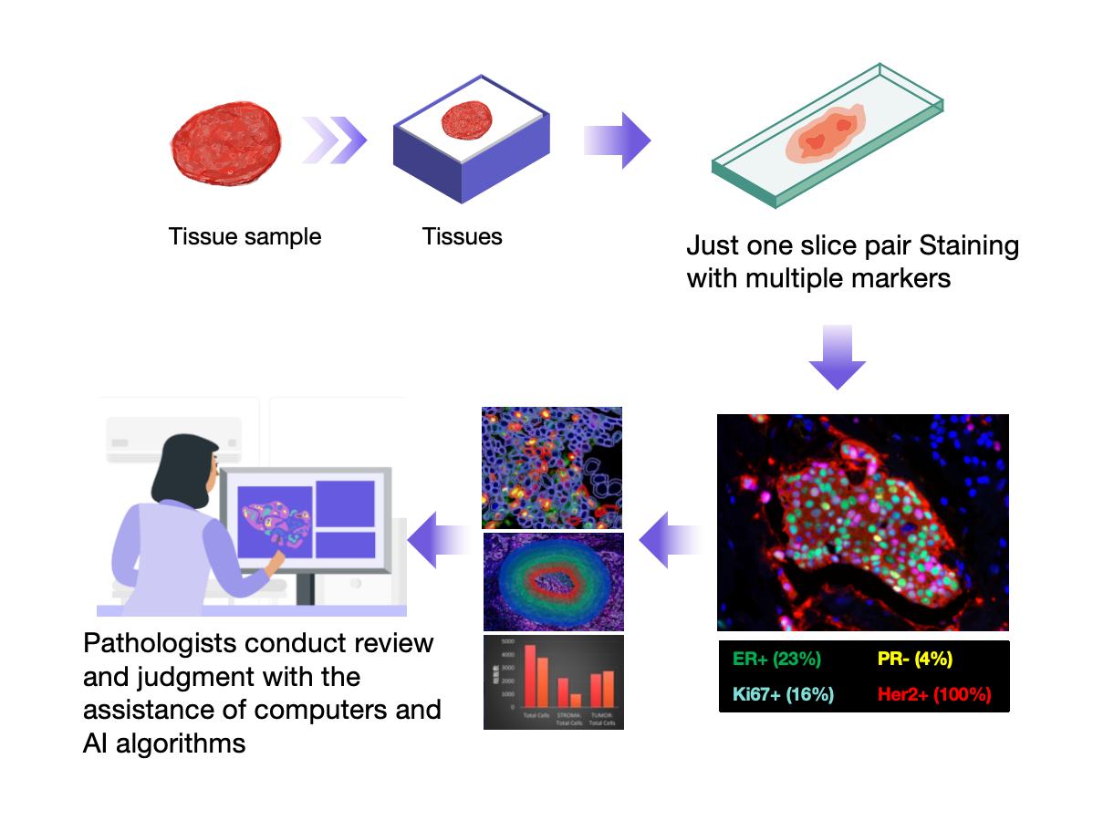
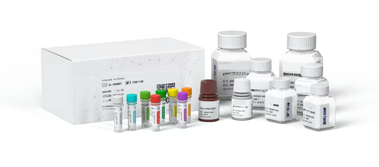
We specialize in the development and manufacture of innovative platforms. The technologies are designed to deliver high-plex, high-throughput spatial profiling of RNA and protein targets directly from a wide range of sample types, including challenging FFPE tissues.
Fortunately, the spectral splitting technology and AI's multi-target hierarchical recognition and analysis capabilities have enabled the entire platform to enter a stage of rapid development, laying the foundation for the eventual application of this technology in heavy daily work. Our advantage lies in the integration of contemporary and professional technologies, making it no longer solely for scientific research use.
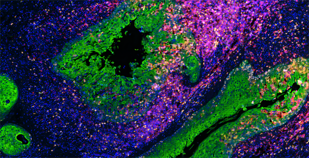
In 2019, we launched our first spatial platform, significantly advancing the ability to visualize multiomic data within the native tissue context. Continuing this trajectory of innovation, our newest FullStack Aurora V8 System, led by Dr. Yuan Mei' research group, featuring the Spatial Molecular Imager, is engineered to provide highly sensitive, sub-cellular resolution.
At HyperLab, we believe that groundbreaking science requires exceptional tools. However, our greatest asset is our team—a dedicated group of scientists, engineers, and professionals driven by ambition, grit, and ingenuity. We are committed to providing our customers with the reliable, high-performance systems needed to answer biology's most challenging questions.